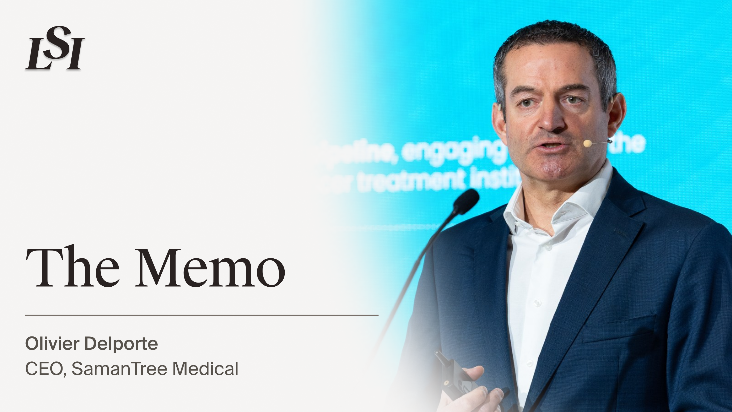
Under the direction of CEO Olivier Delporte, SamanTree Medical is redefining intraoperative tissue imaging with real-time, histology-grade imaging. The company’s flagship device, the HistologⓇ Scanner, uses massively parallel confocal microscopy to capture high-resolution tissue images for surgical margin assessment, at the point of care and without destroying the specimen. Already CE-marked in Europe and FDA-cleared in the U.S., the Histolog Scanner is helping surgeons reduce reoperations and make confident decisions during breast and prostate cancer surgeries. With more than 5,300 patients scanned to date, SamanTree is building a global footprint and expanding into new use cases, from bladder and lung cancers to robotic integration.
Origin Story
SamanTree’s story began at École Polytechnique Fédérale de Lausanne (EPFL), where a multidisciplinary team of engineers and medical innovators set out to solve a high-stakes problem in surgical oncology: the lack of fast, reliable intraoperative margin assessment.
“Too many patients were being brought back for reoperations because surgeons couldn’t confirm the margin status in real time,” said Delporte. “They wanted to eliminate the days or weeks of delay between resection and pathology margin results.”
The team developed a breakthrough imaging modality known as massively parallel confocal microscopy. This innovation formed the foundation of the Histolog Scanner.
Delporte, who joined as CEO after a career bringing impactful medtech innovations to market, said the opportunity was one he couldn’t pass up: “The idea of providing real-time, cellular-resolution imaging in the OR felt like one of those rare moments when technology can immediately improve outcomes and patient experience as well as reduce the impact on the health care system.”
The Current Landscape
SamanTree is tackling the intraoperative margin assessment gap, a critical shortcoming in the standard surgical workflow that leaves patients and their families anxious and uncertain about outcomes and often leads to reoperations. In breast-conserving surgery alone, 15–30%1,2, of patients require a second operation to ensure the cancer is removed. In prostate cancer, the inability to quickly assess margins near neurovascular bundles can lead to avoidable functional side effects such as erectile dysfunction and incontinence3. “Patients deserve better. Too many go home after surgery not knowing if all the cancer was removed.”
The gold standard, frozen section analysis, is accurate but impractical for many settings. It typically takes 45–60 minutes4, requires specialized on-site pathology staff and infrastructure, and involves specimen destruction. “That creates a bottleneck, limiting widespread use,” said Delporte.
Inside the Innovation
“The Histolog Scanner delivers images that allow surgeons and pathologists to achieve equivalent performance to frozen section while reducing turnaround time by 85%5, in approximately one minute per image, with a total turnaround time between eight and 20 minutes,” Delporte explained. The process begins by staining the excised tissue with a fluorescent dye for 10 seconds, followed by rinsing and then placing it in the scanner. Using nearly 30,000 miniaturized lenses, the scanner generates wide-field, high-resolution images of the tissue surface.
“The results are displayed on a touchscreen and can be reviewed collaboratively by the surgical and pathology teams, either on-site or remotely,” said Delporte. “It’s faster, more accessible, and integrates smoothly into the surgical workflow.”
The data speak for themselves. In the prospective SHIELD study6, the use of the Histolog Scanner reduced reoperation rates in breast-conserving surgery by 67% (from 30% to 10%). Clinical performance metrics are strong: more than 90% sensitivity and specificity for margin assessment in breast surgery, and greater than 86% sensitivity and more than 96% specificity in prostatectomy cases.
“We’re the first and only company with massively parallel confocal microscopy,” said Delporte. “We deliver real-time histology-like imaging with no destruction of the specimen at the point of care, a capability no one else offers.”
Progress and Milestones
SamanTree’s momentum is accelerating. Key milestones include:
- FDA 510(k) clearance in Q4 2024, expanding access to the U.S. market
- More than 5,300 patients scanned across multiple cancer types
- Active use at eight U.S. hospitals, with several preparing to become reference centers in 2025–2026
- Strategic U.S. expansion plans, including a possible office and training center by 2026 or 2027
Looking ahead, the company is developing new applications for bladder and lung cancers, real-time biopsy assessment, and AI-assisted image analysis. “We also see opportunities to integrate Histolog into robotic workflows and surgical navigation platforms,” said Delporte.
The LSI Effect
For Delporte, LSI offers the ideal environment to showcase innovation and build strategic momentum. “LSI is where medtech leaders and executives, strategic and financial investors, converge,” he said. “It’s the right place to show what real-time intraoperative imaging can achieve and to find the right investors and partners to bring it to scale in the U.S.”
Join Us at LSI Europe ‘25
Delporte has been selected to present at LSI Europe ’25 (September 7–11) in front of hundreds of global medical technology companies. Join us in welcoming him to the event in London, where he will share the latest updates on SamanTree’s technology and development.
1 Jeevan, R.,et.al. Reoperation rates after breast conserving surgery for breast cancer among women in England: retrospective study of hospital episode statistics. BMJ 2012;345:e4505
2 Kim, Y., et. al. Contemporary Analysis of Reexcision and Conversion to Mastectomy Rates and Associated Healthcare Costs for Women Undergoing Breast-Conserving Surgery. Annals of Surgical Oncology 31, 3649–3660 (2024)
3 Dinneen, E., et al. NeuroSAFE PROOF: NeuroSAFE-guided RARP vs. standard RARP for functional outcomes in localized prostate cancer. The Lancet Oncology. Online First, March 24, 2025. DOI: 10.1016/S1470-2045(25)00091-9.
4 Dinneen E, et al. Intraoperative margin assessment during radical prostatectomy: is microscopy frozen in time or ready for digital defrost? Histopathology. Published online November 1, 2024. doi:10.1111/HIS.15290
5 Baas, J. H., et. al. Confocal laser microscopy for assessment of surgical margins during radical prostatectomy. BJU Int. 2023 Jul;132(1):40-46.
6 Lux MP, Schuller Z, Heimann S, Reichert VMC, Kersting C, Buerger H, Sandor M-F. Re-Operation Rate for Breast Conserving Surgery Using Confocal Histolog Scanner for Intraoperative Margin Assessment—SHIELD Study. Cancers. 2025; 17(10):1640.


© 2026 Life Science Intelligence, Inc., All Rights Reserved. | Privacy Policy | Your Privacy Choices | Delete my Data